X-rays and CT offer predictive power for swine flu diagnosis
June 30, 2009
Diagnostic Imaging.
H.A. Abella
Mexican physicians have compiled a set of radiological findings that is helping local health agencies confirm the diagnosis of the swine A-H1N1 flu virus in humans. Some imaging patterns resemble those from the severe acute respiratory syndrome or ‘avian flu' epidemic that struck mostly Asian countries in 2003.
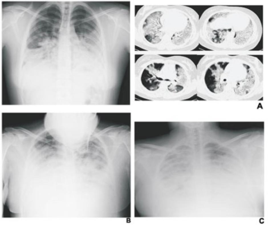
The most outstanding chest x-ray and CT features of the disease include rapidly-progressing basal and axial interstitial/alveolar consolidation and diffuse ground-glass opacities, said Dr. Luis Felipe Alva López, a thoracic radiologist and advisor to Mexico's National Institute of Respiratory Diseases.
Alva López, who is also the current president of the Mexican Society of Radiology, has reviewed most of the Institute's cases and many more at a number of public and private hospitals. He was struck by the similarities and consistencies in radiological patterns between A-H1N1 and SARS.
"When you don't have access to expedite or specific tests, the clinical examination and the chest x-rays could aid the diagnosis in highly suspected patients," he said in response to e-mail questions from Diagnostic Imaging.
The clinical symptoms of swine flu in humans resemble the common seasonal influenza and include fever, lethargy, lack of appetite, coughing, runny nose, sore throat, and in some cases nausea, vomiting, and diarrhea.
To date, the disease has killed 116 people in Mexico alone. About 30,000 cases and more than 100 deaths have been confirmed by the Centers for Disease Control and Prevention in the U.S. The World Health Organization has reported 56,000 people infected worldwide.

CAPTION: Rapidly progressing basal and axial interstitial/alveolar consolidation and diffuse ground-glass opacities are the distinguishing features of swine A-H1N1 flu on chest x-ray and CT. (A) Flu infection in patient 24 hours after clinical diagnosis is shown on x-ray (left) and on CT slices (right). Chest x-rays track patient status 48 (B) and 72 hours (C) after diagnosis (Provided by L.F. Alva López)

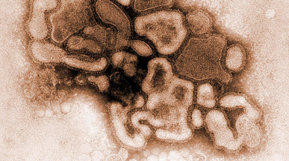
swine A-H1N1 flu virus
ความเห็น (0)
ไม่มีความเห็น